GLP-1 Skin and Hypersensitivity Reactions
GLP-1 drugs trigger skin reactions through receptors in dermal tissue, not just systemic effects.

The distribution of GLP-1 receptors in skin tissue
GLP-1 receptor agonists have become one of the most widely prescribed drug classes in medicine, covering type 2 diabetes, obesity, and obstructive sleep apnea. That scale means dermatologists are seeing these patients constantly now, often without knowing the medication is even in the picture. A cross-sectional analysis published in a dermatology journal notes that dermatology-focused research on cutaneous side effects remains thin, with most of the existing literature limited to individual case reports. That gap has consequences: reactions go unattributed, patients shrug off symptoms as unrelated, and pharmacovigilance databases undercount what's actually happening. This piece works through what's known so far, agent by agent and reaction by reaction, treating the skin as a kind of readout for how well the injection-based delivery model holds up under sustained, real-world use.
Start with where the receptors actually sit. GLP-1 receptors don't stop at the pancreas and brain, the two organs most people associate with this drug class. They also turn up in keratinocytes, dermal fibroblasts, the endothelial cells lining cutaneous microvasculature, and in adipocytes and immune cells living in the skin itself. That distribution matters because it means these drugs can act on skin tissue directly, through a mechanism separate from systemic metabolic change or the physical trauma of a needle going in.
It also sets up a two-sided story, and the two sides don't cancel each other out so much as run on the same wiring. The receptor biology that triggers adverse reactions has, in other contexts, shown signs of benefit: better outcomes in psoriasis, fewer hidradenitis suppurativa flares, improved wound healing. Same pathway, different dose, different tissue, different patient. None of this fully explains any specific rash or nodule discussed later in this piece. But it does explain why the skin's response to GLP-1 signaling isn't some odd coincidence. The tissue is wired to listen, and clinicians who treat the skin as a passive bystander to a drug that works "elsewhere" are missing the point of the receptor map.
Pharmacovigilance data on how often skin reactions are reported
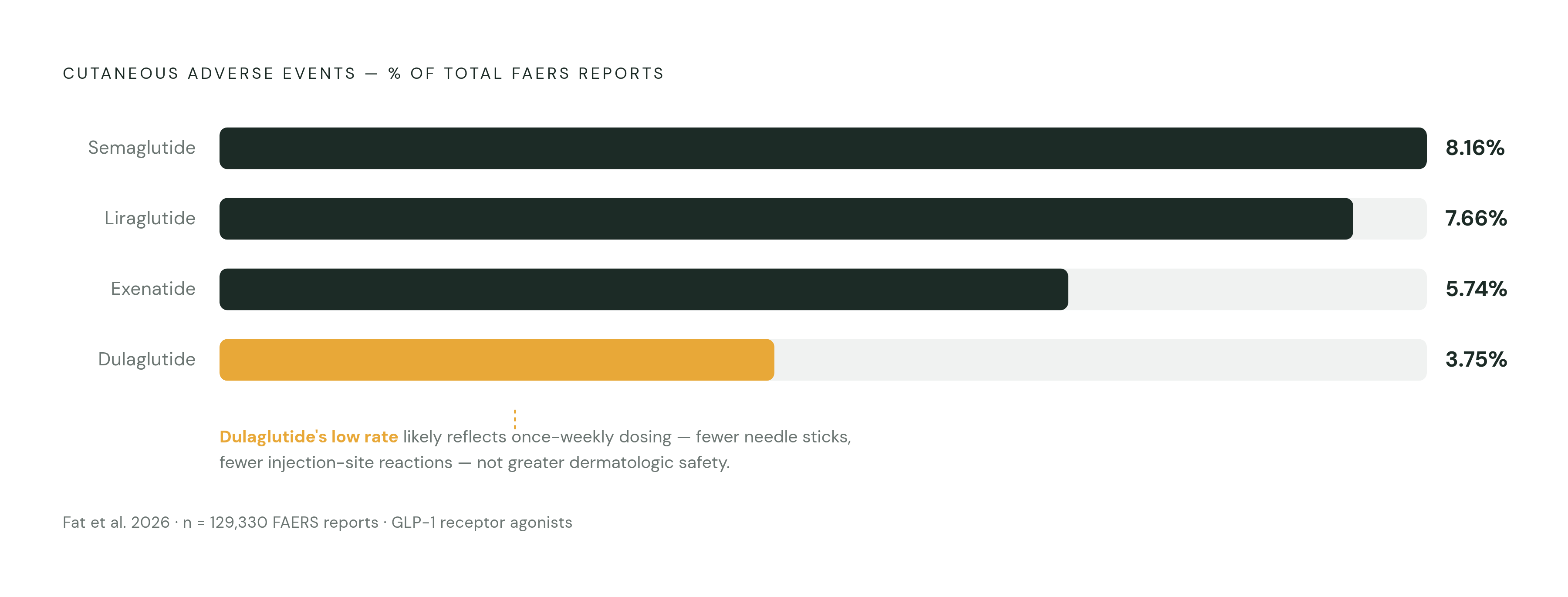
Of 129,330 adverse events reported for GLP-1 receptor agonists in the FDA Adverse Event Reporting System, 5.75% were cutaneous, according to Fat et al. in a dermatology journal, 2026. Of 129,330 adverse events reported for GLP-1 receptor agonists in the FDA Adverse Event Reporting System, 5.75% were cutaneous, according to Fat et al. in a dermatology journal, 2026, and that figure is the aggregate. The real signal appears once the numbers get split by drug, and the split is where most people draw the wrong conclusion.
Semaglutide carried the highest rate of cutaneous cases at 8.16%, followed by liraglutide at 7.66%, then exenatide at 5.74%. Dulaglutide came in lowest at 3.75%, but it also generated the most total adverse events of any agent in the dataset. It would be easy to read that low percentage as dulaglutide being the "safer" skin option. That's the wrong read. The number almost certainly reflects how widely dulaglutide is prescribed rather than any real dermatologic advantage, and Fat and colleagues point instead to its once-weekly dosing schedule as the likelier explanation: fewer needle sticks, less cumulative irritation. That dosing-frequency signal resurfaces later when the injection route itself gets examined directly.
As for what these reactions look like: rash and pruritus dominate the reports, with alopecia and hyperhidrosis trailing behind. Hair loss deserves a second look. It isn't commonly discussed as a GLP-1 side effect, yet it came up repeatedly in the database, per Fat et al., which suggests it's more than background noise.
One caveat has to sit alongside every one of these figures. FAERS is a passive reporting system, nobody is actively screening every patient for a rash and logging it, and what the database actually shows is awareness rather than true incidence. A separate meta-analysis covering 4,861 patients and 396 injection-site reaction events found a risk ratio of 3.55 (95% CI: 2.35 to 5.36) for injection-site reactions with GLP-1 receptor agonists compared to non-GLP-1 comparators, an interval that stays well clear of 1. That interval stays well clear of 1. This is a real, measurable elevation in risk, not statistical noise dressed up as a finding.
Injection-site reactions: the most common cutaneous effect and its underlying drivers
Injection-site reactions are, by a wide margin, the most frequent cutaneous complaint tied to this drug class: localized redness, itching, swelling, sometimes a bit of firmness under the skin where the needle went in. Most resolve on their own, and decent injection technique combined with regular site rotation handles a lot of the problem before it becomes one. That much is uncontroversial.
What's less appreciated is how much of the risk traces back to formulation rather than the drug class as a whole. A 2024 systematic review published in a dermatology journal found that exenatide extended-release accounted for 19 of 33 cutaneous reaction cases, or 57.6% of the total, a lopsided number that has a clear explanation. Extended-release exenatide is delivered as an extended-release preparation that forms a depot under the skin, releasing the drug slowly over time. That depot behaves, in effect, like a foreign object parked in subcutaneous tissue for weeks. A sustained foreign-body stimulus of that kind is the more plausible driver behind both the elevated injection-site reaction rate and, as the next section covers, the eosinophilic panniculitis cases tied almost exclusively to this same formulation. Treat exenatide XR as a different risk category from the rest of the class, distinct from the other entries on a list of interchangeable GLP-1 injectables.
Dosing frequency looks like the one genuinely modifiable lever here. Once-weekly agents cut the cumulative burden of injections on the skin compared to daily dosing, and that's something clinicians can act on now, without waiting on new delivery technology. The stakes go beyond comfort, too: injection-site reactions are a leading reason patients start skipping injections, and in some cases stop treatment altogether. The skin is, in a literal sense, registering where the tolerability limits of this route sit.
Managing this day to day comes down to a short list: rotate sites consistently, pay attention to needle length, let the medication reach room temperature before injecting, and watch for any progression from a mild, self-limited reaction toward something involving deeper subcutaneous tissue.
Rare but serious: eosinophilic panniculitis, bullous pemphigoid, and other severe cutaneous reactions
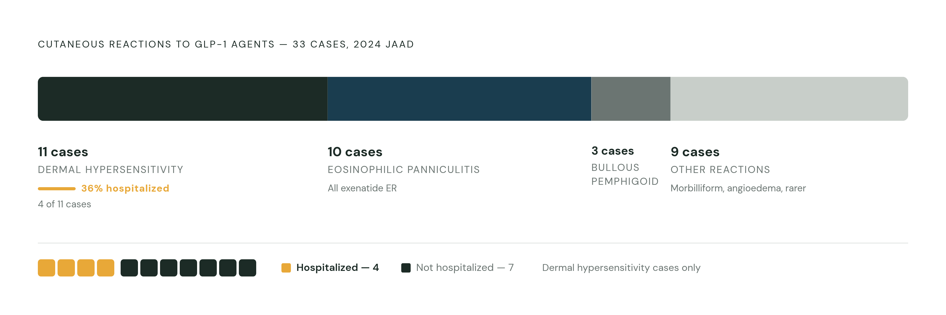
The 2024 JAAD systematic review, presented at the AAD Annual Meeting in San Diego that March, pulled together 31 case reports covering 33 patients. The breakdown matters because the headline shouldn't be the rarity of these events, it should be how badly some of them go.
Dermal hypersensitivity reactions accounted for 11 cases (33.3%): erythematous, itchy wheals and plaques, sometimes localized near the injection site, sometimes spread across the body. Four of those 11 cases, or 36%, required inpatient hospitalization. Treatment ranged from simply stopping the drug to antihistamines, topical or oral steroids, desensitization protocols, and in the more severe cases, intramuscular epinephrine. That 36% figure is the number to actually remember from this section. These aren't reactions that reliably settle down with an antihistamine and a little patience.
Eosinophilic panniculitis made up 10 cases (30.3%): firm, painful, erythematous nodules forming in the subcutaneous tissue near the injection site. Every single one of these 10 cases traced back to exenatide extended-release, which lines up exactly with the depot-formulation mechanism from the previous section. None required hospitalization. Treatment options included stopping the drug, intralesional injections, or surgical excision in more stubborn cases.
Bullous pemphigoid showed up in 3 cases (9.1%), an autoimmune blistering disorder that reflects a process reaching well past the injection site. Its presence in this dataset raises a genuinely open question: can GLP-1 pharmacology trigger immune dysregulation that extends far beyond wherever the needle went in? The case count is small. The mechanism it implies is not.
Rounding out the review: morbilliform drug eruptions (both linked to dulaglutide), angioedema in 2 cases where the GLP-1 agent was considered the most probable cause, and single cases of pyoderma gangrenosum, vesiculopustular dermatosis, psoriasiform drug eruption, and acute photodistributed exanthematous pustulosis. Across all 33 cases, exenatide extended-release accounted for 19, liraglutide for 6, dulaglutide for 5, and subcutaneous semaglutide for 3.
The outcomes offer real reassurance, though. Thirty of the 33 patients recovered completely or were left with only mild post-inflammatory skin changes. Severity here can run high, but it's also largely reversible once the reaction gets recognized and the drug gets stopped or adjusted.
An emerging and underreported symptom: allodynia and skin sensory disturbance with semaglutide
This is the newest thread in the GLP-1 dermatology story, and also the thinnest one, evidence-wise. Allodynia (pain from a stimulus that shouldn't normally hurt) and dysesthesia have been reported as adverse reactions tied to semaglutide, with recognition of this pattern emerging only recently. A recently published case series documented four cases of allodynia tied specifically to dose escalation of semaglutide, not to steady maintenance dosing, and that distinction points clinicians toward the escalation phase itself, despite the small case count, as the period requiring closer attention.
A broader review covering 22 clinical trials and case reports found that oral semaglutide at the 50 mg dose was associated with dysesthesias, hyperesthesia, neuralgia, skin pain, paresthesia, sensitive skin, and a burning sensation on the skin. Incidence for each individual symptom ran roughly 3 to 9 patients out of 334, small numbers on their own, but consistent enough across symptom types to suggest something real rather than coincidental.
What stands out is the timing. These sensory symptoms tracked with dose increases rather than showing up during stable maintenance dosing, which points toward one of two explanations. Either a threshold effect is at work, where higher drug concentrations start directly engaging GLP-1 receptors on cutaneous sensory neurons, or the cause is mechanical: rapid weight loss reshapes subcutaneous tissue and puts tension on nerves that weren't built for that kind of change. Both remain hypotheses, not settled mechanism, and the honest answer is that nobody knows yet which one, if either, is right.
That uncertainty shouldn't translate into dismissal at the bedside. A patient reporting burning, pain to the touch, or unusual skin sensitivity during a dose escalation deserves consideration as a possible drug-related sensory effect, not a shrug. This category, more than any other in this piece, shows how sparse the data really is: no large prospective series exists yet, and the mechanism remains unresolved.
The injection route as a structural contributor to skin risk
Lining up the most severe reactions covered so far makes a pattern visible fast. Eosinophilic panniculitis and dermal hypersensitivity reactions cluster right at the injection site, and eosinophilic panniculitis cases in the JAAD review traced back to exenatide extended-release, the depot formulation that keeps drug sitting in subcutaneous tissue longer than anything else in this class.
Dosing frequency backs this up from a different angle. The FAERS data showed once-weekly dulaglutide with the lowest percentage of cutaneous adverse events (3.75%) despite its wide prescription volume. Cut how often the needle goes in, and skin burden appears to drop, even when the underlying molecule stays roughly the same. That's not a coincidence worth waving off; it's a dose-response relationship between injection frequency and cutaneous harm.
Scale is what turns this from a curiosity into a real exposure problem. The injectable segment held 83% of the GLP-1 market in 2025. The overwhelming majority of patients on this drug class are therefore exposed to whatever dermatologic risk the injection route carries, for as long as they stay on therapy. And therapy, for most patients managing diabetes or obesity, runs for years, not months. Repeated subcutaneous injury at rotating sites, the potential for cumulative immune sensitization to either the excipients or the peptide itself, and the physical presence of depot polymer systems in extended-release formulations: together, these add up to a chronic dermatologic exposure that skin wasn't built to absorb indefinitely.
So how much of what's documented here belongs to the GLP-1 molecule, and how much belongs to subcutaneous delivery as a method? Oral semaglutide's sensory adverse events, the allodynia and dysesthesia from the previous section, suggest the molecule can act on cutaneous biology systemically, with no injection involved at all. But the severe local reactions, panniculitis especially, and injection-site reactions generally, track cleanly with the route of delivery rather than the drug's core pharmacology. One risk belongs to the molecule, and the other belongs to the needle.
The distinction isn't academic. A delivery method that bypasses subcutaneous tissue entirely wouldn't just make injections more pleasant, it would eliminate a whole category of dermatologic risk that the current injection paradigm generates by design. Intranasal delivery, including research into nose-to-brain nanoparticle platforms, is one direction researchers are exploring specifically to sidestep subcutaneous exposure altogether, which would take the injection site out of the equation both as a source of pain and as a site of immune reactivity.
Signs of skin reactions clinicians should watch for and how to respond
Not every rash on a GLP-1 patient carries the same weight. Sorting by severity matters more than treating every cutaneous complaint as one undifferentiated category, and that sorting is where a lot of attribution gets lost.
Mild injection-site reactions, redness, itching, a bit of induration, call for monitoring, a review of injection technique, consistent site rotation, and, where appropriate, a conversation about switching to a less frequent dosing schedule. Urticaria and generalized pruritus deserve closer attention than they usually get. The FAERS data showed more urticaria than expected going in, which raises immune-mechanism questions that warrant real clinical follow-up rather than automatic reassurance.
Firm, painful nodules forming under the skin near an injection site should raise suspicion for eosinophilic panniculitis, confirmed by biopsy. Stopping the drug and intralesional triamcinolone are the standard first-line responses. Widespread or injection-adjacent wheals and plaques point toward dermal hypersensitivity, and given that 36% of these cases in the JAAD review needed inpatient care, the threshold for escalating treatment should stay low, not cautious. Blistering or bullous lesions call for consideration of bullous pemphigoid, a systemic autoimmune condition that needs specialist involvement and prompt action. Burning sensations, allodynia, or pain to the touch, especially around a dose increase, deserve careful documentation and, potentially, a pause on further dose escalation while things are monitored.
Attribution discipline matters throughout all of this. GLP-1 drugs are frequently prescribed alongside other medications, and both the FAERS analysis and the JAAD review make clear that the GLP-1 agent may go unrecognized as a contributing cause. Given how much of the existing literature is limited to individual case reports and passive surveillance, a clinician who notices something unusual and takes the time to document and publish it is doing real work to close a gap that currently sits wide open.
The picture isn't uniformly grim, either. A systematic review by Persson et al., published in Diseases in 2025, looked across 51 studies and found that 17 of them reported beneficial dermatologic outcomes: improvement in psoriasis, fewer hidradenitis suppurativa flares, better wound healing. The dermatology story tied to this drug class runs in both directions at once, and clinical judgment has to hold space for both possibilities rather than defaulting to alarm on one end or dismissal on the other.
Sources
- GLP-1 Agonists Associated With Rare Cutaneous Adverse Reactions
- Injection site reactions with GLP-1 use ‘may be underrecognized’ in clinical practice
- 54813 Rare cutaneous adverse reactions associated with GLP-1 agonists: a review of case reports - Journal of the American Academy of Dermatology
- dermatologytimes.com
- jddonline.com
- medscape.com
- researchgate.net