Nose-to-Brain Drug Delivery Pathway Explained
New imaging study confirms insulin reaches the brain via nasal delivery in humans.

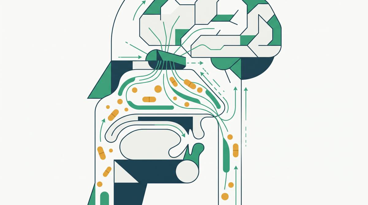
The olfactory epithelium occupies the roof of the nasal passage, posterior to the bridge of the nose. It is specialized sensory tissue, distinct from the respiratory mucosa lining the majority of the nasal cavity. Within it sit olfactory receptor neurons, bipolar cells whose dendrites project into the nasal mucus layer and whose unmyelinated axons fasciculate into bundles that pass upward through the cribriform plate, a perforated section of the ethmoid bone at the base of the skull. Those axons synapse directly in the olfactory bulb, which projects in turn to the hippocampus, amygdala, hypothalamus, piriform cortex, and frontal cortex.
The cribriform plate perforations are where the architecture gets interesting. Nerve fascicles traverse the skull through them, and molecules traveling alongside those fascicles, either within the perineural space or through axonal uptake mechanisms, cross the skull without encountering the blood-brain barrier. No tight endothelial junction to negotiate; no efflux pump to evade. That continuity is precisely what makes this route worth examining seriously.
Sustentacular cells and the lamina propria beneath the epithelial layer provide additional entry points into the perineural compartment. Drug that penetrates to the lamina propria can access the perineural channels surrounding olfactory nerve fascicles, carried toward the CNS by convective flow, a mechanism that operates on a different timescale than axonal transport and one that matters for molecules that cannot efficiently enter neurons.
For years, the mechanistic case for this route rested largely on animal data and theoretical pharmacokinetics. Enough to sustain academic interest; not enough to compel pharmaceutical investment. That shifted with a 2025 PET imaging study using the radiotracer [68Ga]Ga-NOTA-insulin, which provided direct imaging confirmation in living humans that intranasally administered insulin reaches the hippocampus, amygdala, and olfactory cortex within 30 minutes. Insulin is not the therapeutic endpoint. What the study actually accomplished was converting nose-to-brain from an anatomical argument into a measured pharmacokinetic event in the relevant species, the kind of evidence that moves a field from speculative to investigable.
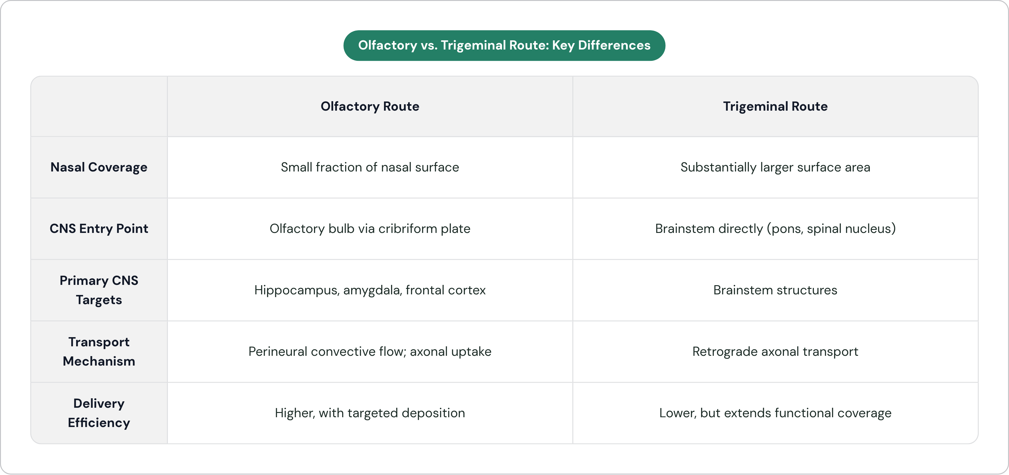
The limitation deserves equal weight. The olfactory epithelium covers only a small fraction of total nasal surface area. Most of the nasal cavity is lined by respiratory mucosa, which has no direct neural connectivity to the olfactory bulb; drug deposited there enters systemic circulation rather than the CNS. That anatomical reality makes targeted deposition to the olfactory region a genuine engineering problem, not an assumption that formulation chemistry alone can satisfy.
The Trigeminal Route: The Secondary Pathway That Extends Coverage Beyond the Olfactory Bulb
The trigeminal nerve, cranial nerve V, innervates the nasal mucosa across a substantially larger surface area than the olfactory epithelium covers. Two of its three divisions are relevant here: the ophthalmic branch (V1) and the maxillary branch (V2) both send fibers into the nasal passages. Trigeminal fibers do not relay through the olfactory bulb. They enter the brainstem directly, connecting to the pons and spinal trigeminal nucleus, which extends down into the cervical spinal cord.
Most delivery discussions move past this distinction too quickly. The olfactory route gives preferential access to limbic and cortical structures: hippocampus, amygdala, olfactory cortex, frontal regions. The trigeminal route runs a parallel path into the brainstem. Together, the two nerve systems mean that N2B delivery can, in principle, reach a wide arc of CNS tissue from a single administration site. But the obvious question lingers: if one route is compromised, say by septal deviation or mucosal inflammation, does the other provide meaningful compensatory coverage, or do both depend on conditions likely to fail together? That question has not been answered in humans with the rigor the clinical application will eventually demand.
Transport along the trigeminal route includes retrograde axonal transport, the same cellular machinery that moves materials from nerve terminals back toward cell bodies. Neurotrophic viruses exploit this pathway to migrate from peripheral tissues into the CNS. That fact should instill both confidence in the mechanism and appropriate humility about what else travels the same road. Molecules taken up at trigeminal nerve endings in the nasal mucosa can migrate retrogradely toward the trigeminal ganglion and brainstem, entering CNS tissue in the process.
Because trigeminal fibers innervate the respiratory mucosa, drug deposited in the lower nasal cavity has a potential CNS-directed route, not only drug reaching the recessed olfactory cleft. Efficiency via the trigeminal pathway is lower than via olfactory deposition, but it extends functional coverage in a way that matters specifically for drugs targeting brainstem structures.
Perineural Channels and the Extracellular Transport Mechanisms That Run Alongside Both Nerve Routes
Nerve fascicles passing through the cribriform plate and along trigeminal branches are surrounded by fluid-filled perineural channels. These are not merely structural compartments. They are dynamic flow conduits. Cerebrospinal fluid pressure gradients drive bulk flow, also called convective transport, along these channels from the nasal submucosa toward brain parenchyma. Molecules dissolved in the perineural fluid move with that flow. The mechanism does not require receptor binding or endocytic machinery; it requires only that the molecule reach the perineural space and remain soluble and intact during transit.
This extracellular mechanism operates in parallel with axonal uptake, and it is particularly relevant for molecules that cannot efficiently enter neurons. Paracellular diffusion, the passage of molecules between epithelial cells through tight junctions, can also contribute, particularly when formulation conditions transiently modulate junction permeability. Certain excipients and absorption enhancers work by loosening tight junctions, though this approach carries mucosal safety considerations that constrain how aggressively it can be deployed.
The distinction between extracellular and intracellular transport carries real consequences for drug design. Small, moderately lipophilic molecules can cross epithelial cell membranes by passive transcellular diffusion and reach the perineural space with relative efficiency. Large hydrophilic molecules, including peptides and proteins, have limited passive permeability and depend on paracellular transport, carrier-mediated uptake, or nanoparticle encapsulation to traverse the epithelial barrier. This is the constraint that determines whether a peptide drug can access the perineural route at all, which is precisely why GLP-1 peptides cannot reach the brain simply by reformulating them as a nasal spray.
Lymphatic drainage alongside perineural channels also governs how long drug remains in brain tissue after arrival. Residence time at the receptor matters, not just initial delivery efficiency.
How Molecules Actually Cross the Nasal Epithelium: The Intracellular Uptake Mechanisms
The nasal epithelium is the first gatekeeping surface any intranasally administered drug must cross. The respiratory region presents a pseudostratified columnar epithelium; the olfactory region presents the specialized neuroepithelium described earlier. Neither is freely permeable to large molecules.
Four primary endocytic pathways mediate intracellular uptake across this surface. Clathrin-mediated endocytosis, the most thoroughly characterized, involves receptor-ligand binding triggering assembly of a clathrin-coated vesicle around the bound molecule. This pathway is saturable and receptor-dependent, so its efficiency is tied to the molecular characteristics of the drug and the receptor expression profile of the target epithelium. Caveolae and lipid raft-mediated internalization operates through membrane lipid domains rather than clathrin, and it is particularly relevant for lipid nanoparticle formulations whose surface chemistry integrates into these domains. Macropinocytosis is a less selective process in which the cell engulfs a volume of extracellular fluid along with any particles suspended in it; it is size-sensitive and less predictable as a targeting mechanism. Receptor- and adsorptive-mediated transcytosis involves a molecule traversing the full epithelial cell and exiting on the basolateral side, facing the perineural space and lamina propria.
Transcytosis is the step that actually matters for CNS delivery. Simple endocytosis moves a molecule into the cell; transcytosis moves it through the cell and deposits it on the other side. For a nanoparticle formulation, surface chemistry can be engineered to favor transcytotic pathways over lysosomal degradation. After endocytic uptake, the fate of the vesicle, whether it traffics to a lysosome or completes transcytosis, is determined in part by the molecular signals presented on the particle surface. That is a design variable, not a biological constant, and it is one of the more underappreciated levers in intranasal peptide delivery.
Two competing processes work against all of this. Mucociliary clearance continuously sweeps nasal contents toward the pharynx; residence time of a droplet in the olfactory region is short unless the formulation includes mucoadhesive components that anchor it there. Enzymatic degradation by peptidases and proteases in the nasal mucosa attacks unprotected peptides on a timescale that can precede meaningful absorption. For peptide drugs, both mechanisms represent active attrition of the available dose before it ever reaches a transporter or endocytic receptor.
Why Nanoparticle Carriers Are Necessary for Peptide Drugs, Not Just Beneficial
The nasal epithelium is not naturally permeable to peptides. GLP-1 and structurally similar peptides are large, hydrophilic molecules for which passive diffusion across epithelial lipid membranes is not a meaningful transport mechanism. They are simultaneously susceptible to enzymatic degradation in the mucosal environment, which means the unencapsulated molecule loses potency before it reaches a transporter or endocytic receptor. A simple aqueous nasal spray formulation of a GLP-1 peptide does not constitute nose-to-brain delivery. It constitutes nasal administration with predominantly peripheral absorption and significant mucosal degradation. That distinction matters when evaluating whether a given formulation approach is worth pursuing at all.
Nanoparticle carriers address several of these problems concurrently. They protect the encapsulated peptide from mucosal enzymes during transit. They can be formulated with mucoadhesive surface chemistries, such as chitosan coatings, that extend residence time at the epithelial surface long enough for meaningful uptake. They engage the endocytic pathways, particularly caveolae-mediated internalization and transcytosis, that cells use for particle uptake. They can also be surface-decorated with targeting ligands directed toward receptors expressed on olfactory neurons or trigeminal nerve endings, and this is where the pharmacokinetic leverage becomes substantive rather than theoretical.
Lactoferrin receptors, for instance, are highly expressed on neurons within the nasal-to-brain pathway. Lactoferrin-decorated nanoparticles have demonstrated substantially greater brain accumulation than unmodified particles at equivalent doses. Surface targeting can meaningfully shift pharmacokinetic distribution toward CNS tissue, not merely nudge it.
The nanocarrier classes in active development for this application span a range of tradeoffs worth examining honestly. Solid lipid nanoparticles and nanostructured lipid carriers offer relatively straightforward manufacturing and established safety profiles; more than 90 research articles were published between 2019 and 2024 specifically addressing these formats for CNS disease treatment via the intranasal route, which reflects both how rapidly this space has grown and how much optimization work remains. PLGA polymeric nanoparticles offer tunable drug release kinetics. Chitosan-based systems bring inherent mucoadhesive properties. Exosomes and biomimetic platforms offer longer-term potential for receptor-level precision but face manufacturing complexity and batch-to-batch variability that limit near-term clinical translation.
Lipid-based formats are favored for near-term clinical development because their safety profile is established across other delivery contexts and their manufacturing processes are scalable. Organizations that have accumulated deep technical depth in precision nanoparticle delivery for peptide encapsulation and nasal delivery, such as Lionbio, which has built more than 30 years of patented intellectual property in this specific space, represent a different category of capability than academic groups demonstrating proof-of-concept in rodents. That distinction bears examination when evaluating which programs are positioned to close the gap between laboratory results and clinical translation.
Deposition Engineering: Why Where a Drug Lands in the Nose Determines Whether It Reaches the Brain
Nasal anatomy creates a deposition problem that formulation chemistry alone cannot solve. The olfactory epithelium is recessed in the upper posterior nasal cavity, anatomically remote from where conventional nasal sprays deposit their payload. Standard spray devices deposit predominantly in the anterior nasal vestibule and lower respiratory mucosa, neither of which connects directly to olfactory neurons. A drug optimally formulated for N2B transport that lands on anterior respiratory mucosa will be absorbed systemically. The biology is sound; the geography defeats it.
Viscosity is one formulation lever for improving olfactory targeting, and the tolerances are narrower than one might expect. A formulation too low in viscosity drains rapidly through the nasal passage, reducing contact time with any epithelial surface. One that is too viscous fails to aerosolize appropriately and does not spread to the recessed olfactory cleft. Wang et al. (2024) demonstrated this relationship with precision: a borneol-modified lipid nanoparticle formulation at approximately 39 millipascal-seconds viscosity achieved an olfactory deposition fraction exceeding a fifth of the administered dose, with a strong correlation to intracerebral drug delivery. Formulations at either higher or lower viscosity performed significantly worse. A single physical parameter, tuned within a tight window, separates meaningful olfactory deposition from systemic absorption. That level of specificity reframes N2B formulation as a disciplined engineering problem rather than a biological hypothesis dressed up as product development.
Device design interacts with these formulation variables in ways the field has historically underappreciated. Spray angle, droplet size distribution, and plume velocity all govern where particles deposit within the turbulent airflow of the nasal passage. Bi-directional delivery devices, which have the patient inhale through the mouth while the device sprays into one nostril, redirect the flow field in a way that preferentially carries particles toward the olfactory cleft. Breath-powered devices reduce the turbulent anterior deposition that conventional pump sprays produce. Neither device innovation works without a matched formulation. Treating formulation and device as sequential decisions rather than a co-design problem is a reliable path to underperformance.
The Limits of the Pathway That Any Serious Delivery Program Must Account For
The absolute fraction of an intranasally administered dose that reaches the brain via direct transport is modest, even under optimized conditions. Whether that fraction produces a pharmacological effect depends entirely on the drug's potency and the receptor density of its CNS targets. High-potency molecules acting at receptors densely expressed in olfactory-adjacent brain regions can produce meaningful effects at brain exposures that would be irrelevant for lower-potency compounds. The question worth asking is not whether N2B delivers large absolute quantities to the brain; it does not. The question is whether what it delivers is sufficient to engage the target, and that calculation is compound-specific.
Interindividual variability in nasal anatomy introduces clinical variability that injectable routes do not face. Septal deviation, mucosal hypertrophy, chronic rhinitis, prior nasal surgery: all alter airflow dynamics and available epithelial surface in ways that affect both deposition and absorption. A clinical population will not have uniform nasal anatomy, and study design must account for that heterogeneity explicitly rather than averaging over it.
Mucociliary clearance rate is not a constant. It varies with respiratory infection, antihistamine use, decongestant use, and general mucosal health. These are not exotic edge cases; they describe routine health states affecting a substantial fraction of any clinical population at any given time. Conditions that slow clearance extend drug residence time; conditions that accelerate it reduce absorption.
The translation problem from animal models to humans is structural and worth stating plainly. Rodent olfactory epithelium covers a proportionally much larger fraction of total nasal surface area than in humans. Positive N2B results in rodent models do not convert linearly to human dose requirements, and assuming they do has historically led to disappointment. Every animal study demonstrating CNS delivery must be interpreted against that anatomical difference, not used uncritically to project human pharmacokinetics.
The field has known about most of these limitations for years. The persistent challenge is designing programs that take them seriously rather than acknowledging them as caveats and moving past them.
Why GLP-1 Peptides Are a Compelling Test Case for the N2B Route — and What the Brain Distribution of GLP-1 Receptors Makes Possible
GLP-1 receptors are expressed in the hypothalamus, hippocampus, brainstem, and reward circuitry. Those regions correspond, with reasonable anatomical precision, to the structures the olfactory and trigeminal routes connect to most directly. The receptor geography and the delivery geography happen to overlap, which makes GLP-1 peptides a structurally coherent test case for N2B delivery in a way that drugs targeting cerebellar structures, for instance, would not be. That alignment is not a given and should not be assumed to hold for other CNS targets without similar mapping.
The injectable GLP-1 market is large. The injectable and parenteral segment held the largest revenue share of a global GLP-1 market valued at USD 66.4 billion in 2025, accounting for nearly 70% of that market. Injectable delivery is effective and commercially validated. From a CNS pharmacology perspective, however, it is indirect: a subcutaneously administered GLP-1 peptide reaches the brain only through systemic circulation, subject to whatever BBB penetration the molecule can achieve in its native or modified form.
The physiological distinction between peripheral and central GLP-1 receptor activation is clinically meaningful. Peripheral receptor activation mediates insulin secretion, gastric motility, and satiety signaling through the vagus nerve. Central receptor activation at hypothalamic circuits mediates direct appetite suppression; at the reward circuitry level, it modulates motivational and hedonic pathways. These are different mechanisms producing overlapping but not identical outcomes. There is also a safety dimension worth examining carefully: the nausea common with injectable GLP-1 therapy is thought to involve overstimulation of receptors at the area postrema, the brainstem emetic center, as a consequence of systemic exposure reaching central emetic circuits at concentrations driven by peripheral dosing requirements.
The N2B hypothesis applied to GLP-1 is that targeted delivery of the peptide to CNS GLP-1 receptors via the nasal route could activate hypothalamic and reward circuitry directly, at lower systemic concentrations, potentially preserving central appetite suppression while reducing peripheral side effects arising from systemic receptor loading. Whether that hypothesis produces a differentiated clinical profile is an empirical question. The mechanism is coherent. But coherence is not validation, and this is where the history of the field counsels skepticism: what gets called nose-to-brain delivery is often just nasal administration, and the two are not the same. Realizing the hypothesis requires the formulation precision, device co-design, and human pharmacokinetic rigor described throughout this piece, not simply an injectable peptide adapted into a spray with the expectation that the biology will cooperate.
The distance between a mechanistically coherent hypothesis and a clinically validated outcome is substantial, and in nose-to-brain delivery, most of that distance is engineering.