GLP-1 Signaling Pathway in Metabolic Regulation
cAMP amplification explains why GLP-1 drugs pose minimal hypoglycemia risk.

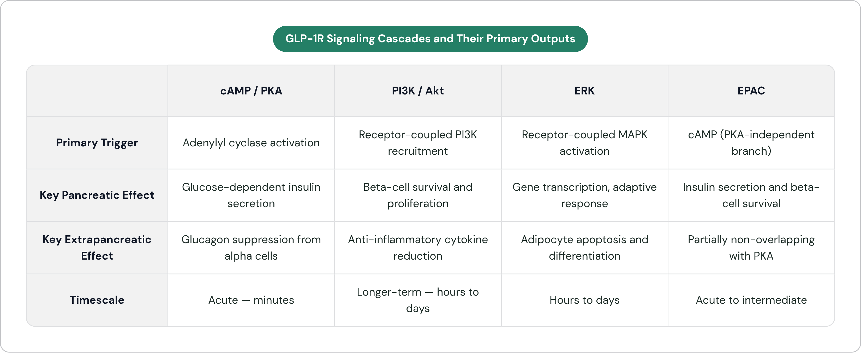
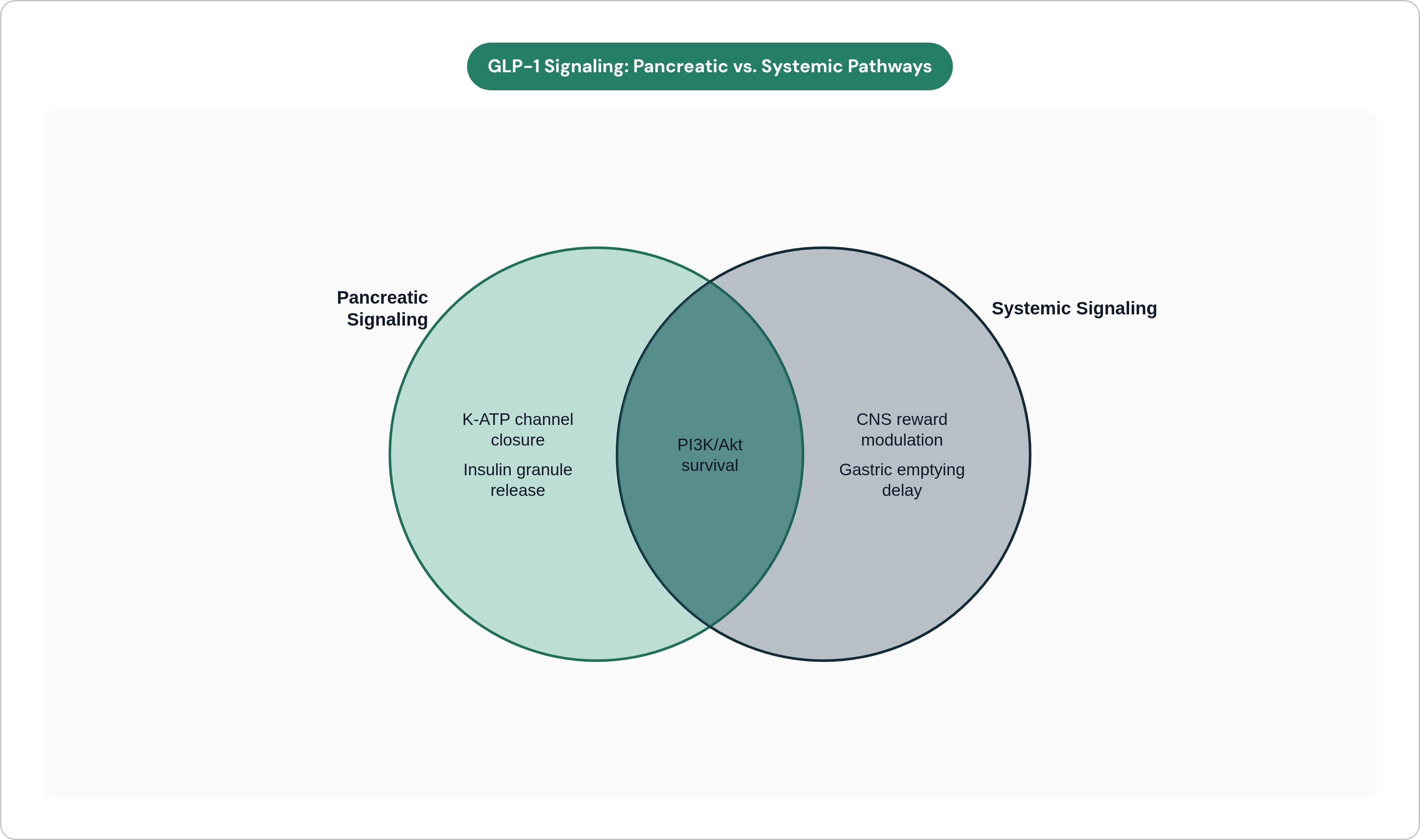
When GLP-1 binds its receptor on a pancreatic beta cell, the proximate biochemical event is activation of adenylyl cyclase, which catalyzes production of cyclic adenosine monophosphate. cAMP is the master signal for most of what GLP-1 does in the pancreas. It activates protein kinase A, which potentiates insulin granule exocytosis and enhances calcium influx. The glucose-derived ATP that closes K-ATP channels and triggers membrane depolarization is already doing its work; GLP-1's cAMP signal amplifies that process rather than replacing it.
GLP-1 receptor agonists carry low intrinsic hypoglycemia risk because their insulin-secreting effect is glucose-dependent: the cAMP amplification they trigger has little to amplify when blood glucose is normal. When GLP-1 binds its receptor, adenylyl cyclase is activated and produces cyclic AMP, which in turn drives protein kinase A to potentiate insulin granule exocytosis and enhance calcium influx. That amplification works on top of the glucose-derived ATP signal that closes K-ATP channels and depolarizes the membrane, it intensifies a process that must already be running, rather than initiating one independently. At normal blood glucose, where K-ATP channel closure and membrane depolarization are not significantly engaged, the cAMP branch has no upstream signal to magnify, so insulin secretion remains near baseline.
Why do GLP-1 receptor agonists carry low intrinsic hypoglycemia risk? Because the cAMP amplification is glucose-dependent, GLP-1R activation intensifies insulin secretion only when glucose is already elevated. Normal blood sugar leaves the amplification signal with little to amplify. But what if that glucose-dependence is not merely a convenient safety property, but actually the central feature of the signaling logic? The low intrinsic hypoglycemia risk of GLP-1 agonists is not a safety feature that was engineered in after the biology was characterized; it falls directly out of that logic.
cAMP also suppresses glucagon release from alpha cells, tightening glucose control from two directions simultaneously. Alongside PKA, cAMP activates EPAC, Exchange Protein directly Activated by cAMP, a PKA-independent branch that contributes to both insulin secretion and beta-cell survival. The cAMP node is not a single-output switch; it distributes through at least two distinct downstream mediators with partially non-overlapping functions.
The PI3K/Akt and ERK Branches That Extend GLP-1's Reach Beyond the Pancreas
The same binding event that triggers cAMP also recruits phosphoinositide 3-kinase, which activates Akt, a kinase with well-characterized roles in cell survival, growth, and metabolic regulation across cell types. In beta cells, PI3K/Akt promotes survival and proliferation, giving GLP-1 a beta-cell-preserving dimension that operates on a different timescale than acute insulin secretion. The cAMP cascade releases insulin now; the PI3K/Akt cascade helps preserve the capacity to release insulin later.
Extracellular signal-regulated kinase activation adds another layer. ERK feeds into gene transcription and cell differentiation, contributing to adaptive responses that unfold over hours and days. In adipose tissue, GLP-1R activates ERK, protein kinase C, and Akt in concert, directly regulating adipocyte apoptosis and preadipocyte proliferation. The fat cell is not simply a passive beneficiary of improved insulin sensitivity; GLP-1R signaling shapes its development and fate at the cellular level.
There are anti-inflammatory dimensions here too. PI3K/Akt modulation reduces infiltrating inflammatory cells in certain tissue contexts and suppresses circulating cytokines including TNF-alpha, IL-1beta, and IL-6. It is also worth considering why these effects recur so consistently across cardiovascular and neurological data — and the answer is that the same molecular logic is operating in those tissues, not because the drug is doing something entirely different in each organ. The signaling looks neatly compartmentalized in a pathway diagram, but in practice the cascades overlap and interact in ways that make clean mechanistic attribution genuinely difficult.
How These Pathways Translate into Effects on Gastric Emptying, Lipid Handling, and Energy Balance
It is worth stepping back from kinase nomenclature to ask what these cascades produce at the organ level.
Gastric emptying slows. GLP-1R signaling in the gut and via vagal afferents delays stomach emptying, blunting postprandial glucose excursions and prolonging satiety. The glycemic curve after a meal is substantially shaped by this effect.
Lipid handling improves. GLP-1 accelerates plasma clearance of triacylglycerol-derived fatty acids and improves insulin signaling in peripheral tissues, reducing ectopic lipid accumulation in liver and muscle, which is itself a driver of insulin resistance.
Thermogenesis is stimulated. GLP-1 activates brown adipose tissue, adding an energy expenditure dimension that operates independently of food intake. The metabolic work continues even when appetite suppression is incomplete.
Adipose tissue composition shifts. By regulating adipocyte apoptosis and preadipocyte proliferation through the cascades described above, GLP-1R signaling influences not just how much fat is stored but the cellular makeup of fat depots and how those depots behave functionally.
None of this is coincidental accumulation. Each effect traces back to the same receptor activating the same set of cascades in different tissue contexts. Once you start thinking about GLP-1's systemic effects as structural features of the receptor's signaling architecture rather than idiosyncrasies of particular drug molecules, the clinical picture becomes considerably less surprising.
Central Nervous System Signaling and the Appetite-Suppression Mechanism
GLP-1R is expressed in hypothalamic nuclei, in brainstem areas including the area postrema and nucleus tractus solitarius, and in reward-related limbic circuits. These populations are not functionally equivalent. Hypothalamic action modulates energy expenditure set points and hunger signaling through neuropeptide release. Brainstem action integrates visceral satiety signals from the gut via the vagus. The reward circuits are where things get genuinely complicated.
Patients on GLP-1 receptor agonists frequently describe a reduction in what they call "food noise": persistent, intrusive preoccupation with food. This phenomenological report has a mechanistic correlate. GLP-1R signaling in mesolimbic circuits modulates the motivational valuation of food, specifically the degree to which the brain's dopaminergic reward machinery assigns salience to food-related cues. This is not aversive conditioning; it is a recalibration of how rewarding food-related cues register. Why exactly does this distinction matter? Because it shapes how we think about durability of effect and patient selection. Nausea is not driving people away from eating.
That shared reward circuit architecture is also why researchers are investigating GLP-1 signaling in substance use disorders. The downstream logic is the same pathway. CNS GLP-1R additionally overlaps with circuits involved in neuroinflammation, mitochondrial function, and protein clearance, which is where the neurodegenerative disease hypotheses originate.
Cardiovascular Signaling: How GLP-1R Activity in the Heart and Vasculature Reduces Cardiac Risk
GLP-1R is expressed in cardiomyocytes, vascular endothelium, and smooth muscle. The receptor is present, the downstream machinery is functional, and the effects are direct rather than simply downstream of improved metabolic control. This is the starting point for understanding why GLP-1 therapeutics have demonstrated cardiovascular benefit.
PI3K/Akt signaling in cardiac cells produces anti-apoptotic effects, protecting cardiomyocytes under stress conditions. Endothelial function improves. Vascular inflammation is reduced through the same cytokine-suppression mechanisms operative in peripheral metabolic tissue. Mitochondrial function in cardiac tissue is enhanced, reducing oxidative stress in an organ that runs continuously at high energy demand and is correspondingly sensitive to oxidative damage.
GLP-1 receptor agonist treatment is associated with reductions in major adverse cardiovascular events in clinical trial data, a signal that cannot be fully explained by weight loss. One might argue that cardiovascular benefit is simply a consequence of losing fat mass — but that argument undersells the direct vascular and myocardial biology and may generate incorrect predictions about which patients benefit and under what conditions. This is a clinically consequential distinction, not a theoretical one.
The direct cardiac signaling also helps explain why cardiovascular outcome trials became central to GLP-1 drug development even when the primary indication was glycemic control. The biology was pointing somewhere before the clinical evidence confirmed it.
Neurological Indications: What GLP-1 Signaling Suggests About Dementia and Addiction
Type 2 diabetes substantially elevates the risk of Alzheimer's disease. The shared pathophysiology is instructive: insulin resistance in the brain, chronic neuroinflammation, oxidative stress, mitochondrial dysfunction, and impaired protein clearance all contribute to both conditions. GLP-1R signaling addresses each of these through mechanisms already described in peripheral tissues. The question is whether those mechanisms are operative in the brain at the concentrations that systemic administration actually achieves.
The neuroprotective case is mechanistically coherent. GLP-1 receptor agonists reduce alpha-synuclein aggregation via enhanced autophagy, modulate microglial-driven neuroinflammation, and support dopaminergic neuron survival through anti-apoptotic signaling. Phase 2b data from the ELAD trial, presented at AAIC 2024, showed that liraglutide reduced brain volume loss across frontal, temporal, parietal, and total gray matter regions compared to placebo.
Then the EVOKE trials reported in late 2025: the largest GLP-1 trial ever conducted in Alzheimer's disease, testing semaglutide, missed its primary endpoint. That raises an important question worth sitting with rather than explaining away. The mechanistic rationale was sound. A phase 2 signal existed. The phase 3 result did not follow. That gap is not a clean refutation of the underlying biology; phase 3 failures in Alzheimer's trials have a long history of reflecting trial design problems as much as target problems. But it does raise a hard question about whether the limiting variable was CNS drug exposure, patient selection, disease stage, or something else. I do not think the field has a confident answer yet, and I am skeptical of anyone who claims otherwise.
For addiction, early clinical and preclinical data across alcohol, opioid, and stimulant use disorders suggest that GLP-1R signaling in mesolimbic and mesocortical circuits modulates drug reward through mechanisms that parallel its effects on food reward. The receptor distribution overlaps substantially; the transduction logic is the same. Whether current delivery approaches can access these circuits reliably enough to produce durable clinical benefit remains an open question.
Biased Agonism and the Next Generation of Pathway-Selective GLP-1 Drugs
Different ligands engaging different regions of GLP-1R can preferentially stabilize receptor conformations that favor particular downstream partners. This is biased agonism: not simply activating or inhibiting the receptor, but selectively weighting which intracellular pathways it preferentially engages.
The practically relevant distinction is between cAMP/PKA pathway activation, which drives most of the established therapeutic benefit, and beta-arrestin recruitment, which drives receptor internalization and is associated with certain side-effect profiles. Biased agonists designed to favor cAMP signaling over beta-arrestin show longer-lasting glucose reduction, greater food intake suppression, and more pronounced weight loss in research settings. The therapeutic window widens not by adjusting dose but by changing the conformational preference of receptor engagement.
Multi-receptor agonists represent a parallel strategy. Co-targeting GLP-1R alongside receptors for gastric inhibitory polypeptide and glucagon combines complementary signaling cascades for additive or synergistic metabolic effects. The signaling architecture is not a ceiling; it is a design space, and the field is still mapping its parameters.
This is also where delivery platform innovation becomes scientifically relevant in a non-trivial way. Reaching specific receptor populations in specific tissues, brain versus gut versus adipose, with the right agonist profile is partly a chemistry problem and partly a delivery problem, and the two cannot be cleanly separated.
Why GLP-1's Broad Reach Makes Delivery Route a First-Order Scientific Question
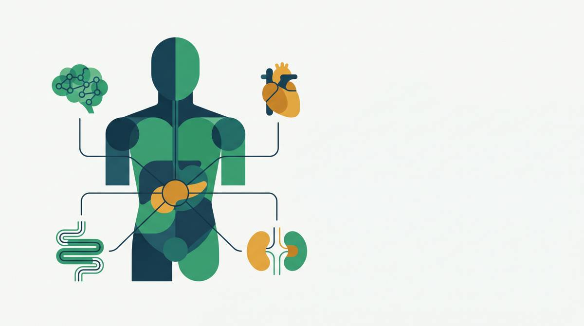
The same receptor, activated in different tissues, produces categorically different functional outputs: insulin secretion in pancreatic beta cells, appetite suppression in hypothalamic neurons, oxidative stress reduction in cardiomyocytes, reward circuit modulation in mesolimbic structures. Tissue context determines what the signal means. But how does this affect our original premise about GLP-1 as a systemic therapeutic? It means that where a GLP-1 agonist distributes in the body is not a secondary question about pharmacokinetics; it is a primary question about what the drug is actually doing.
Injectable administration distributes a GLP-1 receptor agonist systemically, reaching most receptor populations without tissue specificity. CNS penetration is limited by the blood-brain barrier, though circumventricular organs lacking a complete barrier provide partial access. Gastrointestinal side effects, nausea, vomiting, abdominal pain, affect a substantial proportion of patients on current formulations, and mechanistically this tracks with high systemic concentrations hitting gut GLP-1R broadly. Real-world persistence data compound the concern: a large proportion of patients discontinue GLP-1 therapy within the first year, with tolerability among the most frequently cited reasons.
For neurological indications, the delivery problem sharpens further. The EVOKE result, considered alongside the broader pattern of mechanistically credible hypotheses failing in CNS disease trials, invites serious attention to whether insufficient drug concentration in relevant brain compartments is a rate-limiting variable. This is the scientific context in which intranasal delivery platforms designed to exploit olfactory and trigeminal pathways become pertinent, not as a convenience formulation, but as a potential prerequisite for achieving therapeutic concentrations in reward and neurodegeneration-relevant circuits. Some nose-to-brain nanoparticle platforms take this approach, designed to load CNS compartments preferentially via these pathways and potentially alter both tissue distribution and the tolerability profile associated with systemic administration. Lionbio, a nasal-spray peptide biotech built on Columbia University nanoparticle IP, is one example pursuing this route for GLP-1 delivery.
What I have come to believe, slowly and with some resistance, is that GLP-1 therapeutics will advance through better agonist design and through more precise delivery working together. The signaling architecture is capable of producing remarkable effects. Getting the right signal to the right tissue at the right concentration is the problem the field actually needs to solve, and it has not solved it yet.