Obesity-Brain Axis and Hypothalamic Inflammation
Inflammation in the brain's appetite center may explain why obesity resists conventional treatment.
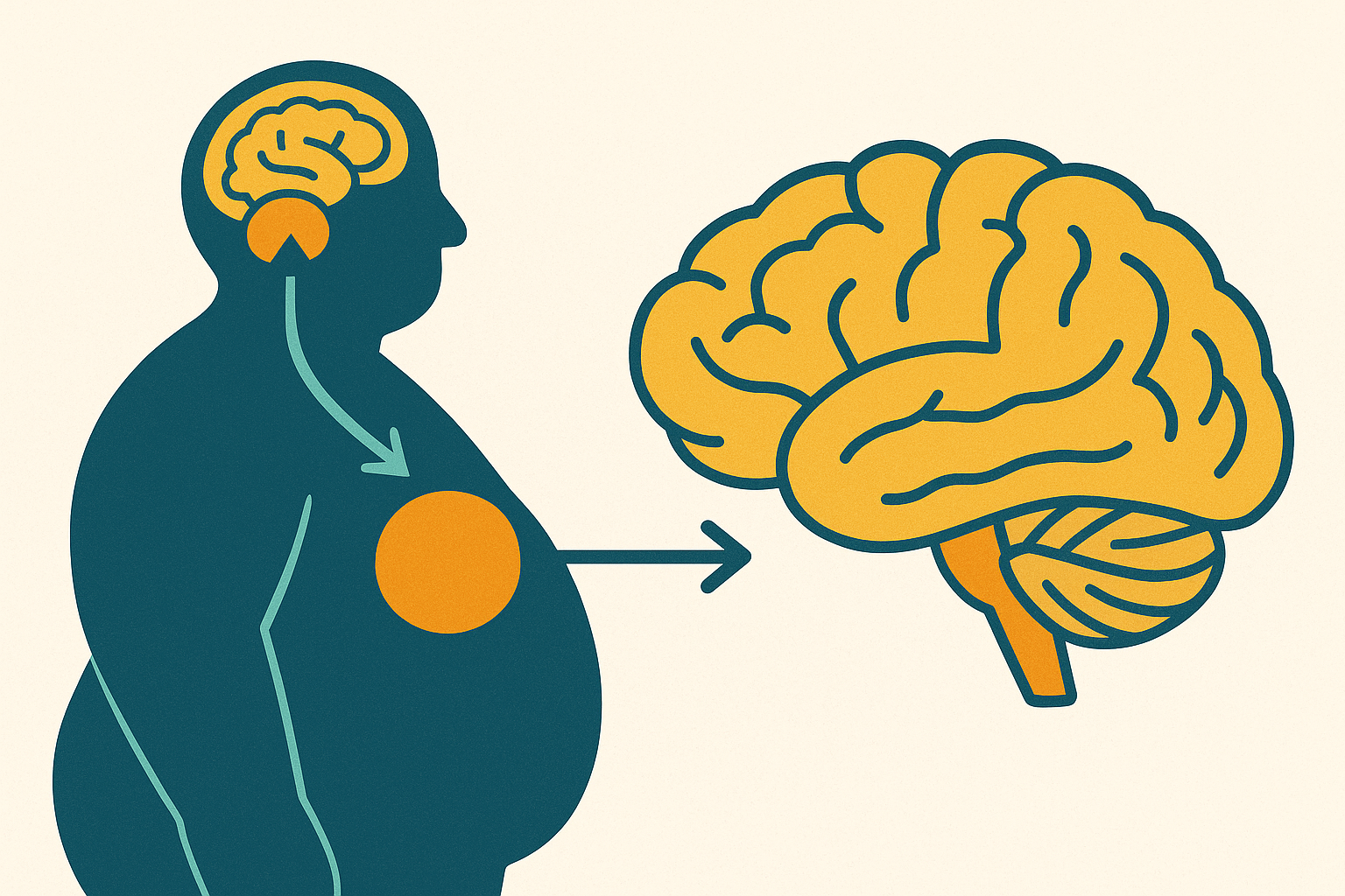
Obesity has long been framed as a matter of willpower or bad math on calories in, calories out, but the data increasingly point to something else: a disease of chronic inflammation inside the hypothalamus, the brain region running the circuitry that tells us when to eat and when to stop. Take that seriously and it changes what an effective treatment actually has to do, and it changes where you'd even start looking for one.
For decades the public conversation stayed fixed on energy balance. Eat less, move more, let the numbers sort themselves out; it's a tidy model, and it isn't wrong exactly, but it treats the brain like a passive scorekeeper tallying inputs and outputs. The hypothalamus is setting the rules of the game, not just keeping score. It takes in a constant stream of peripheral signals (leptin from fat tissue, insulin from the pancreas, GLP-1 from the gut, adiponectin from adipose cells) and turns that information into moment-to-moment decisions about hunger, fullness, and how many calories the body burns at rest.
Two structures do most of the work here. The arcuate nucleus, or ARC, functions like a switchboard, home to two neuron populations pulling in opposite directions: AgRP/NPY neurons driving hunger, POMC neurons signaling satiety. The paraventricular nucleus, or PVN, takes the ARC's output and translates it into downstream effects on appetite and energy expenditure. When the circuit works, it self-corrects: eat enough, leptin rises, POMC activates, and hunger fades. Fifteen years of obesity research has complicated that picture considerably, though, showing the hypothalamus behaving less like a lone conductor and more like an ecosystem with its own internal politics. Astrocytes, microglia, tanycytes: these were once filed under structural filler, cells that just held the neurons in place. Turns out they're active participants in metabolic regulation, and a command center where the support staff has gone rogue explains, far better than diet alone ever could, why weight loss so rarely holds.
How chronic overnutrition triggers inflammation inside the hypothalamus
Here's the part that hasn't fully landed in public conversation: in animal models, hypothalamic inflammation shows up before obesity does. Diets high in saturated fat activate innate immune pathways inside hypothalamic tissue within days, before any measurable weight gain registers on a scale. The sequence, then, runs differently than assumed: the inflammatory trigger arrives first, riding in on the fat content of the diet itself, and the metabolic fallout follows behind it, sometimes weeks behind it.
Microglia respond first. They're the brain's resident immune sentinels, and in a healthy hypothalamus they mostly patrol, clearing debris, keeping things quiet in the background. Chronic exposure to high-calorie, high-fat signaling pushes them toward a pro-inflammatory state, one that dumps cytokines like TNF-α, IL-1β, and IL-6 straight into the neural environment around the ARC. Those cytokines don't stay put, interfering with the signaling machinery hunger and satiety neurons depend on, adding static to a channel that needs to stay clear.
Astrocytes follow next. Reactive astrocytes proliferate around ARC neurons in a process called gliosis, and that word is worth pausing on, because it sounds like scarring, like an inert aftermath, something that just sits there once the damage is done. The reality runs the opposite direction: the astrocyte proliferation actively remodels the synaptic architecture POMC and AgRP neurons rely on to talk to each other, so the scarring becomes a mechanism of disease rather than a leftover symptom of one.
Then come the tanycytes, a cell type that deserves more attention than it gets. They line the third ventricle and act as gatekeepers, deciding which peripheral hormones actually reach the ARC, and inflammation weakens that barrier function. Once the gate loosens, the ARC's exposure to circulating signals gets less controlled, feeding right back into the same inflammatory loop that damaged the barrier to begin with. Impaired neurons allow more caloric intake; more caloric intake drives more inflammation; more inflammation impairs more neurons further. There's no obvious exit built into that loop, which is part of what makes this disease so stubborn.
For years this whole picture rested on rodent studies. Fair question to ask: does any of it actually happen in people, or is this another case of mouse biology that doesn't translate? Recent work using advanced MRI to assess hypothalamic microarchitecture has started answering that, picking up signs consistent with inflammation and gliosis in the hypothalami of adults and children with obesity alike. That's a real bridge, moving hypothalamic inflammation out of the rodent lab and into something you can see, in living human tissue, on a scan.
Leptin resistance as the signature failure mode of inflamed hypothalamic circuitry
Leptin is supposed to make the whole system work. Fat cells secrete it roughly in proportion to fat mass, so as fat stores grow, leptin rises, and that rise signals energy sufficiency at the ARC, suppressing NPY and activating POMC. Mechanistically, leptin binds its receptor, activates JAK2, and sets off a STAT3 phosphorylation cascade that changes gene transcription in ways that reduce appetite. On paper, it's an elegant loop, closed and self-correcting.
Obesity produces a paradox anyone who's struggled with weight recognizes instinctively, even without knowing the biochemistry: people with obesity typically run high circulating leptin, not low, and yet the hunger doesn't go away. That's resistance, by definition, a system awash in the satiety signal that still can't hear it. Inflammation gives a direct answer for why. Chronic hypothalamic inflammation upregulates a protein called SOCS3, which blocks the JAK2 step in leptin's signaling cascade. Layer on endoplasmic reticulum stress inside ARC neurons and glial remodeling of the synapses those neurons depend on, and the leptin receptor stops transmitting even when leptin is sitting right there in abundance, practically shouting.
Adiponectin tells a parallel story that gets a lot less airtime, oddly, given how much it matters. Its receptors, AdipoR1 and AdipoR2, show up not just in the hypothalamus but across the cortex, thalamus, and hippocampus, and not just on neurons but on microglia and astrocytes too. Adiponectin normally supports neuronal integrity and helps keep neuroimmune activity in check. In obesity, levels tend to drop, pulling out a stabilizing influence at exactly the moment the hypothalamic immune environment needs it most.
The clinical weight of this is hard to overstate. Leptin resistance reflects a breakdown in the brain's own fullness signal, one where the receiving circuitry can't hear a message that keeps getting sent. There's no natural correction once gliosis sets in: more fat mass produces more leptin, more leptin exposure deepens the resistance, and the inflammatory backdrop against which all of this plays out keeps intensifying. It works like a ratchet, one direction only, with nothing built in to release it.
The broader network: how the gut, adipose tissue, and inflamed brain communicate
None of this happens in isolation inside the skull. The hypothalamus sits in the middle of a signaling network running continuously between gut, fat tissue, and brain, and the traffic runs both ways rather than flowing down from some central authority barking orders.
GLP-1 is the clearest example of gut-to-brain signaling. Released from the intestine after a meal, it reaches the brain by two routes: part of it crosses the blood-brain barrier directly to act on receptors in the PVN and ARC, and a second signal travels indirectly, activating vagal afferent nerves that relay up to the nucleus tractus solitarius in the brainstem before reaching hypothalamic targets. Adipose tissue contributes leptin and adiponectin, as already discussed, but in obesity, fat tissue itself becomes inflamed and starts dumping its own pro-inflammatory cytokines into systemic circulation, some of which appear able to cross into brain tissue. The liver may contribute signals of its own too, though the evidence here is newer and causality in humans is still being worked out. Better to say that plainly than pretend the case is closed.
Layered on top of this homeostatic system is a separate, hedonic one: the reward circuitry built around mesolimbic dopamine that drives food-seeking independent of actual energy need. Anyone who's ordered dessert after a full meal has felt the hedonic system override the homeostatic one firsthand, no biochemistry degree required. Obesity appears to dysregulate both at once, and an inflamed hypothalamus, one that can't exert normal homeostatic suppression anymore, leaves the hedonic drive to eat comparatively unchecked. The two failures compound rather than simply add.
Some of this likely starts earlier than adulthood, before years of diet and environment have had a chance to act at all. Prenatal exposure to maternal obesity or diabetes has been identified as a risk factor for hypothalamic inflammation and gliosis in offspring, along with elevated obesity risk later in life, pushing the origin point of this disease back before birth in at least some cases. Chronic stress, poverty, endocrine-disrupting chemicals, and adverse childhood experiences have all been implicated as contributors to hypothalamic gliosis too, a reminder that this disease gets shaped by circumstance as much as by diet in the narrow sense. And the downstream associations keep expanding: hypothalamic inflammation and gliosis have been linked to reproductive disorders including PCOS and male hypogonadism, with investigators actively exploring possible ties to metabolic liver disease and sarcopenia. The obesity-brain axis keeps turning up in places researchers didn't expect to find it.
Why peripheral GLP-1 therapies work — and where their limits lie
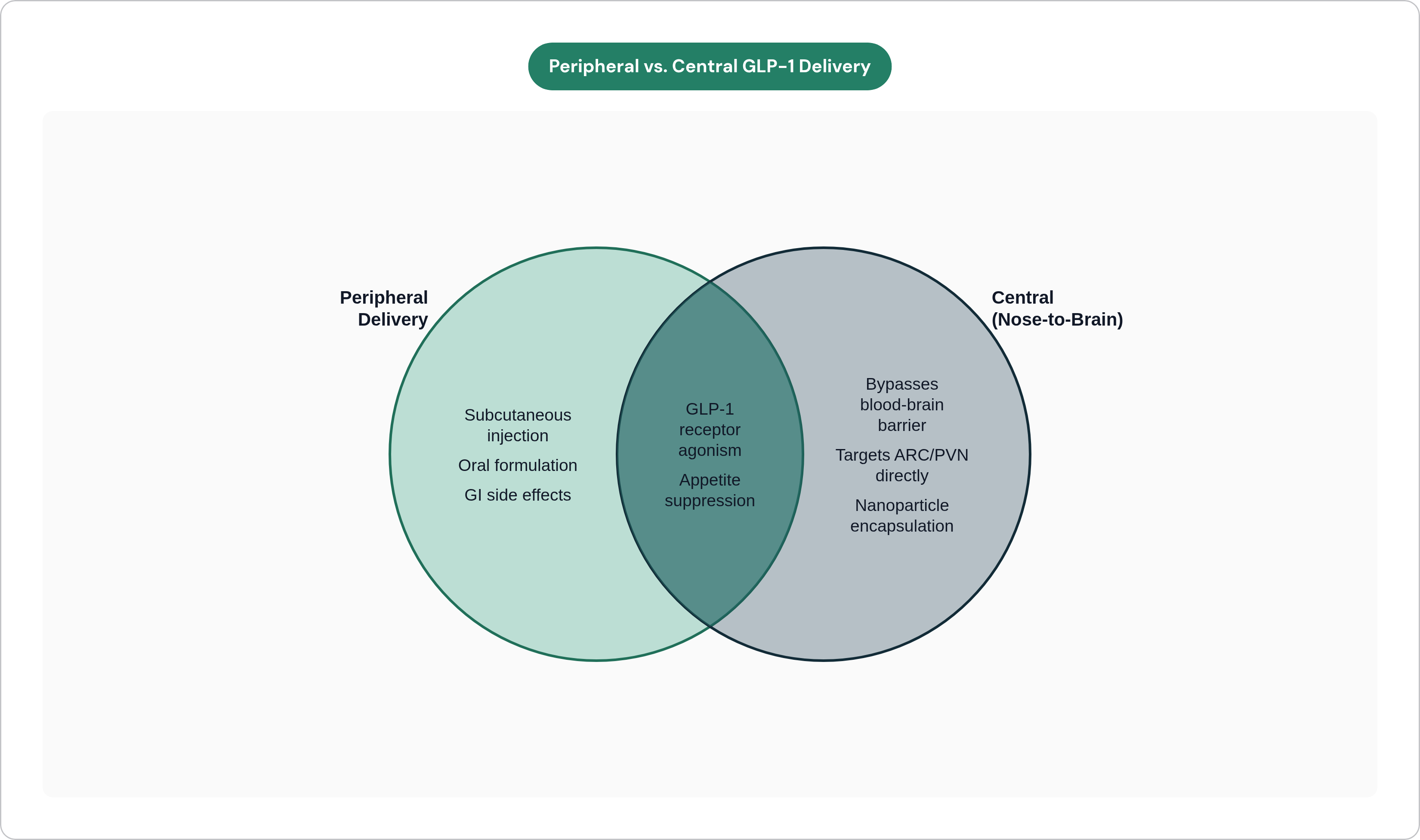
GLP-1 receptor agonists work by taking a signal the brain already understands and stretching it out, holding it at pharmacological levels the body would never produce on its own. Endogenous GLP-1 already reaches ARC and PVN receptors through the two routes described above; therapeutic agonists mimic that signal but sustain it over hours instead of the brief post-meal window natural GLP-1 offers. The result is appetite suppression, slower gastric emptying, and better insulin dynamics, and the effect shows up clearly at population scale. GLP-1 therapies posted roughly $132 billion in global sales in 2025, up 33.5% year-over-year, with the weight-loss indication specifically growing at a 131% five-year compound annual rate. That's not a small signal, and the underlying biology works — the market is telling you so in numbers that are hard to argue with.
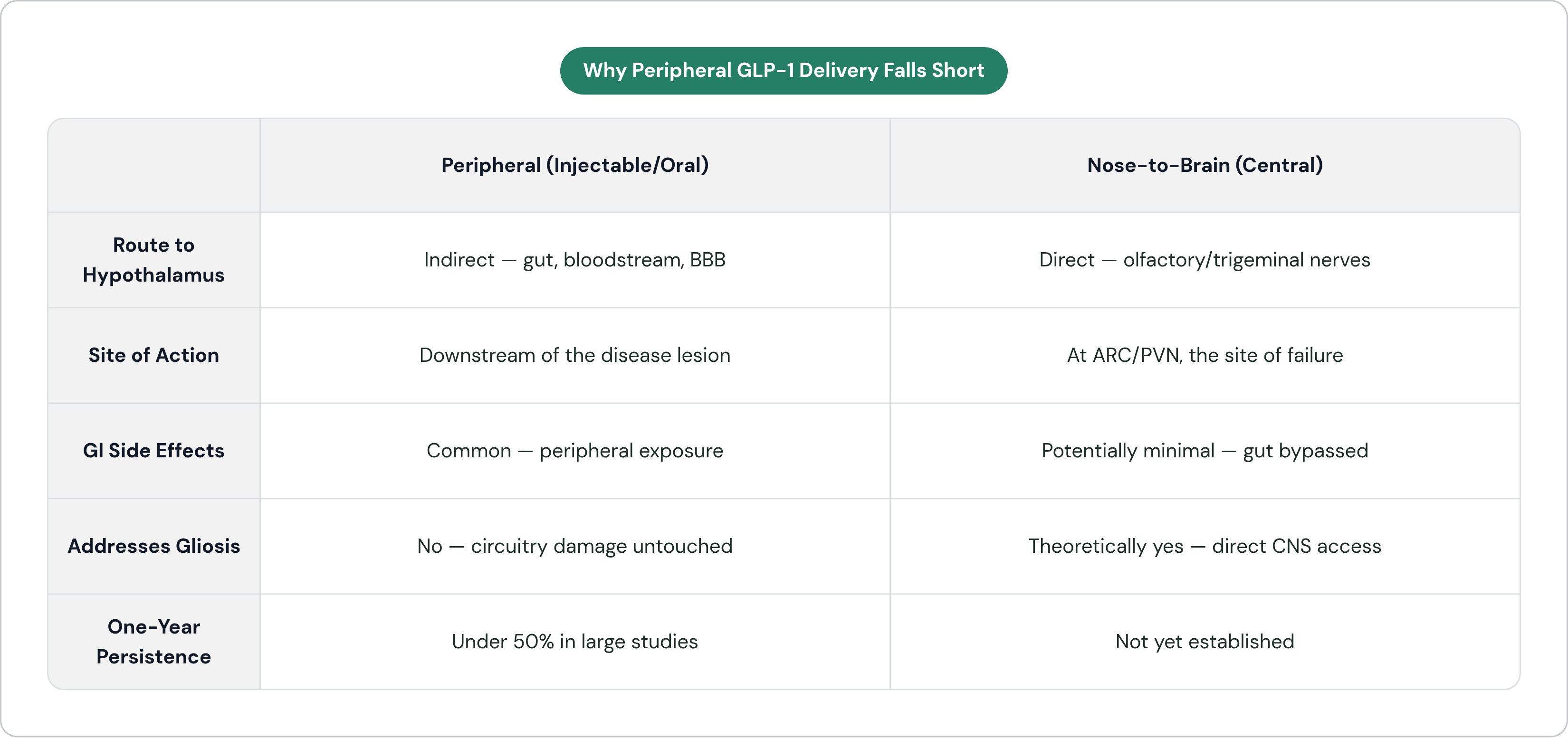
So how does this square with the original problem, the inflamed hypothalamus itself? Peripheral delivery, whether by injection or, increasingly, an oral pill, means the drug reaches its central targets indirectly, diluted by everything it passes through first: gut, bloodstream, liver, blood-brain barrier. Much of the nausea, vomiting, and gastroparesis tied to GLP-1 therapy traces back to the drug acting in the periphery before it ever reaches the hypothalamus. Which suggests the side-effect profile comes from the delivery route, not from some unavoidable property of the molecule itself. Add the physical burden of a lifetime of subcutaneous injections, and the persistence numbers start to make more sense.
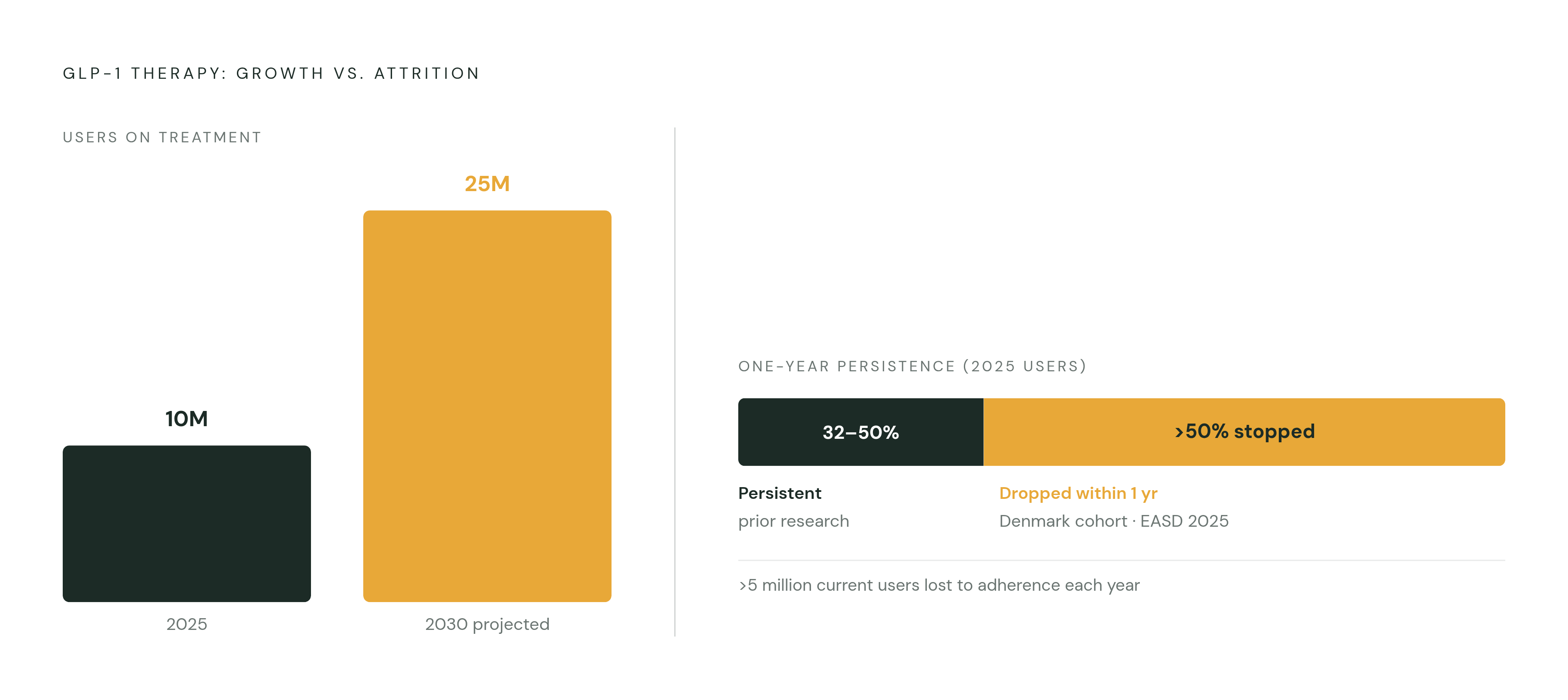
And the numbers are sobering, frankly. Prior research put one-year persistence on GLP-1 therapy somewhere between 32% and 50%. A large population-based study out of Denmark, presented at the 2025 European Association for the Study of Diabetes meeting, found that more than half of GLP-1 receptor agonist users had stopped treatment within a year. Extension data from the STEP trials shows what happens next: patients who quit tend to regain the weight, often most of it. Oral formulations cut the needle burden, which matters to plenty of patients, but they don't touch the GI side effects, and they don't improve how precisely the drug reaches its CNS targets. What peripheral delivery leaves untouched, in every formulation on the market right now, is the actual site of disease: the gliosis, the leptin resistance, the disrupted ARC circuitry sitting inside the hypothalamus itself.
What it would mean to deliver GLP-1 directly to the inflamed hypothalamus
That raises an obvious question, one that seems almost too simple once you say it out loud. If the disease lives in the hypothalamus, why does treatment start in the gut, or under the skin?
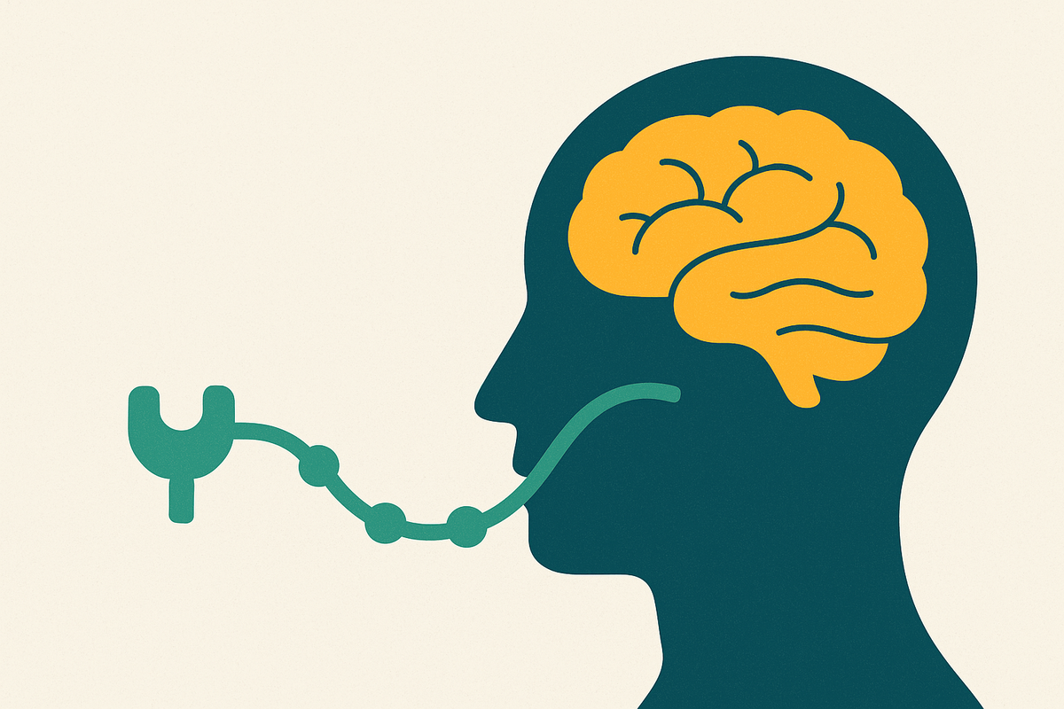
There's an anatomical route that offers a different answer: nose to brain. The olfactory and trigeminal nerve pathways run a direct physical conduit from the nasal epithelium into the central nervous system, bypassing both the blood-brain barrier and systemic circulation entirely. A drug deposited along these pathways can, in principle, reach brain tissue without ever touching the gut, the liver, or the peripheral vasculature, which happen to be the exact tissues responsible for most of GLP-1's characteristic side effects.
The engineering here is harder than it sounds, though. Peptides don't survive the nasal environment unprotected; enzymatic activity and mucociliary clearance in the nasal epithelium degrade unformulated peptides well before they reach neural tissue. That's why nanoparticle encapsulation matters here, shielding the peptide from enzymatic breakdown while improving mucosal adhesion and uptake into the nerve-associated channels leading into the CNS.
Follow the mechanistic logic and it holds together. A drug that reaches the ARC and PVN at meaningful, sustained concentrations engages the exact receptors whose signaling gliosis and leptin resistance disrupted in the first place. Relying on a peripheral cascade to relay a message through an already-compromised relay system carries a built-in ceiling; central delivery could act right at the point of failure instead. Some platforms have been built around this idea, drawing on more than 30 years of patented nanoparticle delivery IP originating at Columbia University, purpose-built to protect and transport peptides along the nose-to-brain route. The technology grew out of foundational academic science and has since been adapted for this specific delivery method.
The platform implications reach beyond GLP-1 and beyond obesity, too. The same delivery architecture built to get a peptide past the nasal barrier and into hypothalamic circuits could, in principle, carry other neurologically active peptides relevant to addiction or neurodegeneration, conditions where a subcutaneous injection has never been a particularly good fit for reaching the brain in the first place.
What remains unknown here is real, and it deserves saying outright instead of glossed over with a hedge. Central delivery of GLP-1 agonists is young science, and whether it actually cuts GI side effects in practice remains an open question. Whether centrally-mediated anti-inflammatory effects differ meaningfully from peripheral ones, how dose-response shifts with direct CNS access: these are open questions the field is still chewing on. That kind of honesty matters more here than in most corners of drug development, precisely because the mechanistic claims on the table are bold ones.
Reframing what it means to treat obesity effectively
If hypothalamic gliosis and disrupted ARC circuitry sit at the root of this disease, durable remission probably requires treating those central lesions directly, not just managing the metabolic symptoms downstream of them. Chronic appetite suppression through a peripheral drug that most patients stop taking within a year functions more like a workaround than a cure, a fragile one at that.
The adherence numbers read almost like a design brief for what has to come next. Roughly 10 million Americans were on GLP-1 treatment in 2025, with projections putting that near 25 million by 2030. Yet the gap between people who start treatment and people still on it at the one-year mark represents millions cycling back toward weight regain and the metabolic risk that comes with it. That gap isn't a footnote; it's the central design problem the next generation of obesity treatment has to solve.
Worth remembering, too, that hypothalamic gliosis has social and environmental roots: stress, poverty, endocrine disruption among them. No delivery technology, however precise, replaces the need for broader public health work on those upstream drivers. Better drug delivery enables better treatment, but it doesn't answer the deeper question of why so many people develop hypothalamic inflammation in the first place.
Where does the science point from here? Toward anti-inflammatory strategies aimed at hypothalamic gliosis itself, with appetite suppression treated as a downstream proxy rather than a stand-in for the real target. Toward delivery technologies that can reach CNS targets with real precision while cutting the peripheral burden driving both side effects and dropout. And toward an expanding set of neurological indications, addiction and dementia among them, where obesity-brain axis research keeps surfacing shared pathophysiology with other disorders of the hypothalamus and limbic system.
The GLP-1 market, at its current scale, is evidence the underlying biology works: a drug that mimics a natural satiety signal produces real, measurable weight loss in real people. The adherence crisis sitting right next to that success is its own kind of evidence, though, and it points the other way, suggesting the delivery paradigm hasn't caught up to where the disease actually lives. Biology flagged the brain as the site of disease years ago, and the therapies are still finding their way there.